
Knee
Normal Anatomy of the Knee Joint
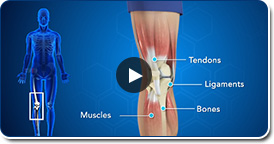
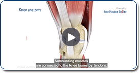
The knee is made up of four bones. The femur or thighbone is the bone connecting the hip to the knee. The tibia or shinbone connects the knee to the ankle. The patella (kneecap) is the small bone in front of the knee and rides on the knee joint as the knee bends. The fibula is a shorter and thinner bone running parallel to the tibia on its outside. The joint acts like a hinge but with some rotation.
ACL Hamstring
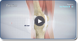
Anterior cruciate ligament (ACL) reconstruction hamstring method is a surgical procedure that replaces the injured ACL with a hamstring tendon. Anterior cruciate ligament is one of the four major ligaments of the knee that connects the femur (thigh bone) to the tibia (shin bone) and helps stabilize your knee joint.
Meniscus Tear
Meniscus tear is one of the most common knee injury in athletes, especially those involved in contact sports. A sudden bend or twist in your knee cause the meniscus to tear. These injuries are generally associated with knee pain and swelling.
Patellofemoral Instability
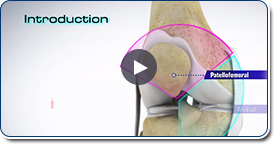
The knee can be divided into three compartments or spaces patellofemoral, medial and lateral. The patellofemoral compartment is the compartment in the front of the knee, and may become unstable after injury or due to the alignment of the limb. The medial compartment is the area on the inside portion of the knee, and the lateral compartment is the area on the outside portion of the knee joint.
Arthroscopy of the Knee Joint
Knee Arthroscopy is a common surgical procedure performed using an arthroscope, a viewing instrument, to look into the knee joint to diagnose or treat knee problems. It is a relatively safe procedure and the majority of patients are discharged from the hospital on the same day of surgery.
ACL Patellar Tendon
Anterior cruciate ligament (ACL) reconstruction patellar tendon is a surgical procedure that replaces the injured ACL with a patellar tendon. Anterior cruciate ligament is one of the four major ligaments of the knee that connects the femur (thigh bone) to the tibia (shin bone) and helps stabilize the knee joint.
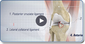
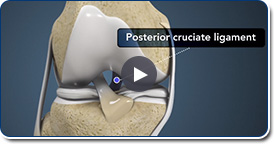
Posterior Cruciate Reconstruction
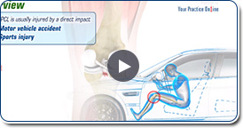
Posterior cruciate ligament (PCL), one of four major ligaments of the knee are situated at the back of the knee. It connects the thighbone (femur) to the shinbone (tibia). The PCL limits the backward motion of the shinbone. PCL injuries are very rare and are difficult to detect than other knee ligament injuries. Cartilage injuries, bone bruises, and ligament injuries often occur in combination with PCL injuries. Injuries to the PCL can be graded as I, II or III depending on the severity of injury.
Patellofemoral Pain Syndrome
Patellofemoral pain syndrome, also referred to as PFPS, is one of the most commonly reported knee problems, accounting for one in four knee complaints seen by orthopedists.
Osteochondral Autograft
Osteochondral autografting (OCG) is a surgery to repair damaged articular cartilage that lines the ends of bones in a joint. An osteochondral autograft is a piece of tissue taken from a healthy section of the joint and transplanted to replace the chondral defects in the joint. There are two techniques used in osteochondral autografting: mosaicplasty and osteochondral autograft transfer system (OATS).
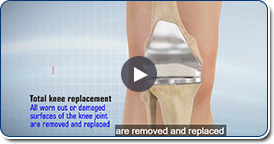
Total Knee Replacement
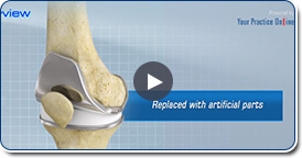
Total knee replacement, also called total knee arthroplasty, is a surgical procedure in which the worn out or damaged surfaces of the knee joint are removed and replaced with artificial parts. The knee is made up of the femur (thigh bone), the tibia (shin bone), and patella (kneecap). The meniscus, the soft cartilage between the femur and tibia, serves as a cushion and helps absorb shock during motion.
Medial Collateral Reconstruction
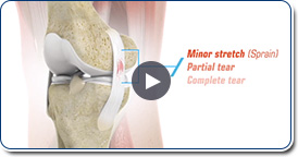
Medial collateral ligament (MCL) is one of four major ligaments of the knee that connects the femur (thigh bone) to the tibia (shin bone) and is present on the inside of the knee joint. This ligament helps stabilize the knee. An injury to the MCL may occur as a result of direct impact to the knee. An MCL injury can result in a minor stretch (sprain) or a partial or complete tear of the ligament.
Micro Fracture

Microfracture is a surgical technique used to repair articular cartilage damage in the knee called chondral defects. Articular cartilage is a complex avascular (no blood supply) tissue which consists of cells called chondrocytes suspended in a collagenous matrix. It appears as a smooth, shiny, white tissue at the ends of the bones which come in contact with each other to form a joint.
Multiligament Reconstruction
The knee is the most complex joint in the body and is formed by the articulation between the thigh bone (femur) and the shinbone (tibia). A knee cap is present over the front of the joint to provide extra protection. These bones are held together by four strong rope like structures called ligaments. Two collateral ligaments are present on either side of the knee and control the sideway movements of the knee.
Osteochondral Allograft

An osteochondral allograft is a piece of tissue taken from a diseased donor to replace damaged cartilage that lines the ends of bones in a joint. A section of cartilage and bone is removed, shaped to precisely fit the defect and then transplanted to reconstruct the damage.
Meniscal Transplant
Meniscal transplantation is a surgical procedure to replace the damaged meniscus of the knee with healthy cartilage. The meniscus is a C-shaped cartilage ring that acts like a cushion between the shinbone and the thighbone. Each of your knees has two menisci - one on the inside (medial aspect) and the other on the outside (lateral aspect) of your knee.
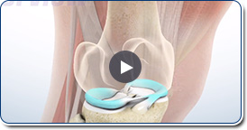
Subchondroplasty
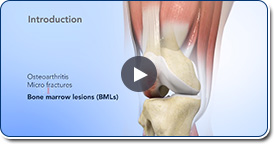
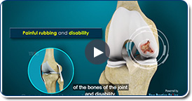
Knee osteoarthritis (OA) is a common form of arthritis that causes joint pain and stiffness. It is a progressive disease in which the joint cartilage gradually wears away and may lead to disability. Bone marrow lesions or BMLs are strong predictors of osteoarthritic cartilage damage. Bone marrow lesions are visible on an MRI but not in a regular X-ray.
Uni Knee Replacement
Unicompartmental knee replacement is a minimally invasive surgery in which only the damaged compartment of the knee is replaced with an implant. It is also called a partial knee replacement. The knee can be divided into three compartments: patellofemoral, the compartment in front of the knee between the knee cap and thigh bone, medial compartment, on the inside portion of the knee, and lateral compartment which is the area on the outside portion of the knee joint.
Knee fracture
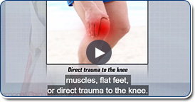
A fracture is a condition in which there is break in the continuity of the bone. In younger individuals, these fractures are caused from high energy injuries, as from a motor vehicle accident. In older people, as the bones become weaker, fractures most commonly occur with slips and falls.