
HIP
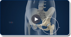
Normal Anatomy of the Hip joint
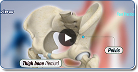
The hip joint is the largest weight-bearing joint in the human body. It is also referred to as a ball and socket joint and is surrounded by muscles, ligaments, and tendons. The thigh bone or femur and the pelvis join to form the hip joint. Any injury or disease of the hip will adversely affect the joint's range of motion and ability to bear weight.
Hip Arthroscopy
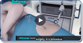
Hip arthroscopy, also referred to as keyhole or minimally invasive surgery, is a procedure in which an arthroscope is inserted into a joint to evaluate and treat various joint injuries. An arthroscope is a small, fiber-optic instrument consisting of a lens, light source, and video camera. The camera projects an image of the inside of the joint onto a large screen monitor allowing the surgeon to look for any damage, assess the type of injury, and repair the problem.
Labral Tears
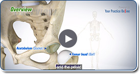
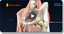
A hip labral tear is an injury to the labrum, the cartilage that surrounds the outside rim of your hip joint socket. The hip joint is a ball and socket joint in which the head of the femur is the ball and the pelvic acetabulum forms the socket.
Femoro Acetabular Impingement (FAI)
Femoroacetabular impingement (FAI) is a condition where there is too much friction in the hip joint from bony irregularities causing pain and decreased range of hip motion. The femoral head and acetabulum rub against each other causing pain and damage to the hip joint.
Hip Chondral Injuries
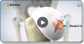
The hip joint is one of the largest weight-bearing joints in the body, formed by the thigh bone or femur and the acetabulum of the pelvis. It is a ball and socket joint with the head of the femur as the ball and the pelvic acetabulum forming the socket.
Hip Bursitis
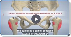
Hip bursitis is a painful condition caused by inflammation of a bursa in the hip. Bursae are fluid filled sacs present in joints between bone and soft tissue to reduce friction and provide cushioning during movement. The bony prominence of the hip is called greater trochanter and is present on the outer side of the upper thigh bone or femur. The bursa overlying it is called trochanteric bursa.
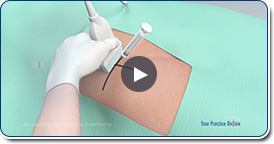
X-ray Guided Hip Injection
Hip joint injections involve injecting medicine directly into the hip joint to diagnose the source of pain or treat pain due to conditions such as arthritis, injury or mechanical stress of the hip joint. Hip pain may be experienced in the hip, buttock, leg or low back.
Gluteus Medius Tear
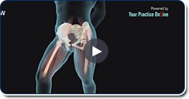
A gluteus medius tear is a condition characterized by severe strain on the gluteus medius muscle that results in partial or complete rupture of the muscle. The gluteus medius is one of the major muscles of the hip and is essential for movement of the lower body and keeping the pelvis level during ambulation.
Ultrasound Guided Hip Injection
The hip joint is a ball and socket joint and is the largest weight-bearing joint in the human body. The head of the thigh bone or femur forms the “ball” and the acetabulum of the pelvis is the “socket”. These bones come together to form the hip joint.